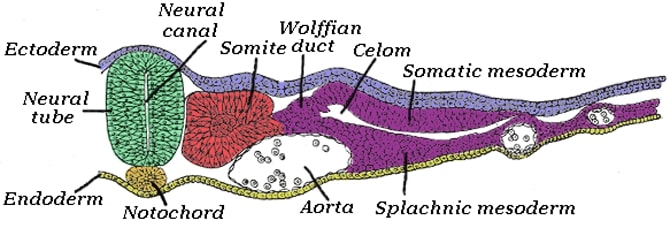
The muscular system develops from the mesodermal germ layer, with the exception of part of the smooth muscle. It includes skeletal muscle, smooth muscle found in various parts of the body, and cardiac muscle.
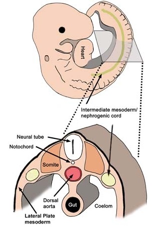
Skeletal muscle differentiates from the paraxial mesoderm, which forms somites in the occipital and sacral regions, as well as somitomeres in the cephalic region.
Smooth muscle is formed from the splanchnic mesoderm surrounding the intestine and its appendages, as well as from the ectoderm in certain areas such as the pupils, mammary glands, and sweat glands.
Cardiac muscle is formed from the splanchnic mesoderm surrounding the cardiac tube.
The first sign of muscle cell differentiation is the elongation of cells that will become myoblasts. The first smooth muscle to appear is in the esophagus wall during the fifth week of development.
In the third month of development, the cells of striated muscles differentiate into myofibrils, which consist of clear and dark bands. These bands are limited by bundles of fibrils.
The formation of motor plates occurs when nerve fibers contact myoblasts.
The musculature of the axial skeleton, limbs, walls of the torso, and head is formed from somites and somitomeres. Somites form and differentiate from the occipital region and continue caudally. They give rise to the sclerotome, dermatome, and the two regions from which the musculature will form.
In the process of forming limb and trunk wall musculature, cells in the ventro-lateral margin zone contribute to the formation of the myotome and provide progenitor cells. Cells from the dorso-medial margin of the dermatomyotome migrate ventrally towards the future dermatome. They also contribute to the formation of the myotome and give rise to the posterior trunk musculature.

During differentiation, myoblasts, the precursor cells, fuse and form long, multinucleated muscle fibers. Myofibrils appear in their cytoplasm, and by the end of the third month, transverse striations that give the skeletal musculature its characteristic appearance become visible.
A similar process occurs in the seven pairs of somitomeres located in the cephalic region, rostral to the occipital somites. Somitomeres have a reduced organization and do not give rise to the sclerotome and dermatomyotome.
Tendons, which support muscle insertion on bones, are formed from the sclerotome cells located adjacent to the myotomes at the anterior and posterior margins of the somites. The development of these cells is regulated by the transcription factor scleraxis (SCX), which also plays a role in tendon healing in adults.

The distribution of musculature is influenced by the connective tissue into which myoblasts migrate. In the cephalic region, connective tissue develops from neural crest cells. In the cervical and occipital regions, connective tissue is derived from somitic mesoderm. In the trunk and limbs, connective tissue is derived from somatic mesoderm.
During the fifth week of development, muscle cells form two structures: the epimere and the hypomere. The epimere, a smaller dorsal structure, is composed of cells from the dorso-medial region of the somites. These cells reorganize and form the myotomes. On the other hand, the hypomere, a larger ventral structure, is formed by the migration of cells from the dorso-lateral region of the somites.
The muscle segments are innervated by nerves that have two branches: a primary dorsal branch for the epimere and a primary ventral branch for the hypomere. The corresponding nerves will continue to innervate the same muscle segment throughout the downward migration process.
The myoblasts of the epimere form the extensor muscles of the vertebral column, while the myoblasts of the hypomere form the muscles of the limbs and trunk wall. The cervical portion of the hypomere gives rise to the prevertebral muscles, scalenes, and geniohyoid muscles.
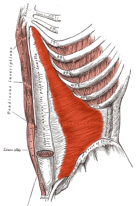
In the thoracic segments, the myoblasts separate into three layers, which become the external intercostal, internal intercostal, and transversus thoracis muscles in the thoracic wall. In the abdominal wall, these three muscle layers form the external oblique, internal oblique, and transversus abdominis (Tra) muscles.
The muscles corresponding to the thoracic wall maintain their segmental character due to the presence of ribs. However, the muscles corresponding to different segments of the abdominal wall fuse and form extensive muscle structures.
The myoblasts from the lumbar segments generate the formation of the quadratus lumborum muscle, while the myoblasts from the sacral and coccygeal regions form the pelvic diaphragm and the striated muscles of the anus.
At the ventral end of the hypomeres, a ventral longitudinal muscle column is formed. In the abdominal region, this column is represented by the rectus abdominis muscle, while in the cervical region, it is represented by the infrahyoid muscles.
The muscles in the cephalic region under voluntary control, such as the muscles of the tongue, eyeballs, and pharyngeal arches, are derived from the paraxial mesoderm. The distribution of these muscles is controlled by connective tissue derived from neural crest cells.
The muscles of the head and trunk, except those derived from the branchial arches, derive from myotomes. In the 8 mm embryo, the myotomes fuse on the surface, giving the trunk wall a segmented appearance. The intercostal muscles remain segmented, while the dorsal muscle mass remains unique.
In the third month of development, the rectus abdominal muscles differentiate and grow towards the midline, leading to the appearance of the linea alba.

In the seventh week, the limb muscles begin to form as a condensation of mesenchyme near the limb primordium (the limb buds). The distribution of these muscles is controlled by connective tissue derived from the somatic mesoderm, which also forms the bones of the limbs.
Initially, the limb muscles have a segmented structure, but during development, the segments fuse, resulting in muscles containing tissue from multiple segments.
The upper limb primordia are located on both sides of the last five cervical segments and the first two thoracic segments. The lower limb primordia are located on both sides of the last four lumbar segments and the first two sacral segments.
After the formation of the primordia, the corresponding spinal nerves penetrate into the mesenchyme. The ventral and dorsal branches of these nerves fuse to form large ventral and dorsal nerves.
The spinal nerves play an important role in the differentiation and motor innervation of the limb muscles, as well as providing sensory innervation to the dermatomes. The orderly arrangement of the dermatomes can be identified in adults, although their initial disposition changes with the growth of the extremities.
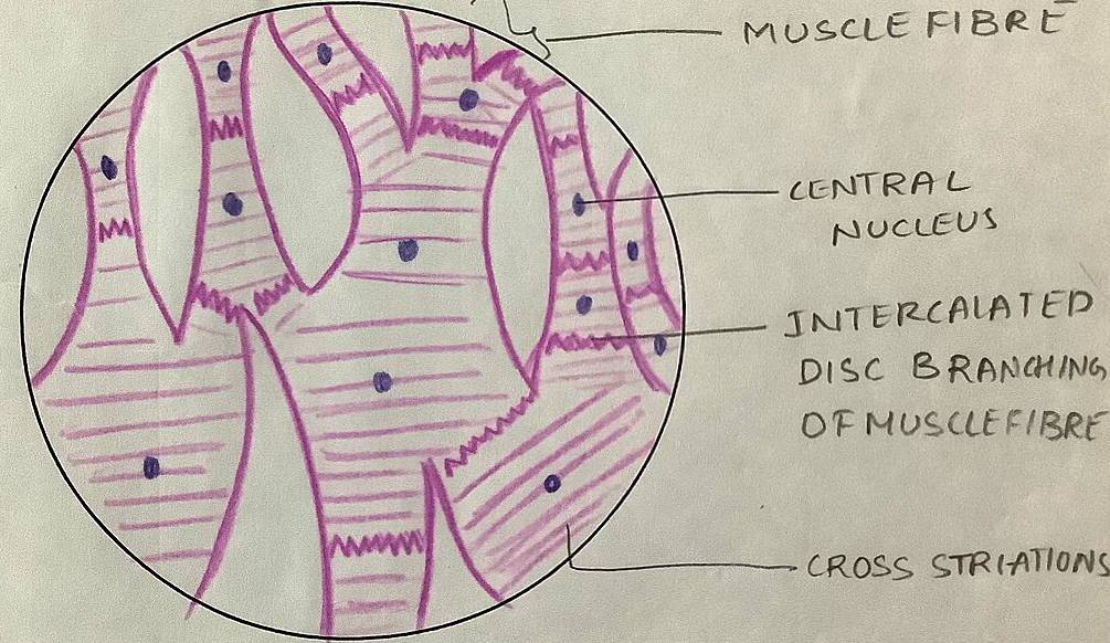
The cardiac muscle, or myocardium, is derived from the splanchnic mesoderm that surrounds the endothelial cardiac tube. Myoblasts connect with each other through special structures that later give rise, in the final period of fetal life, to the intercalated discs characteristic of this type of musculature. Similar to skeletal muscle, myofibrils undergo the same developmental process, but myoblasts do not fuse. In advanced stages of development, several bundles of special muscle cells become visible, known as Purkinje fibers, which have irregularly distributed myofibrils and make up the heart's conduction system.

Smooth musculature in different parts of the body is derived from various embryonic origins. The dorsal aorta and major arteries have smooth muscle cells that come from the mesoderm of the lateral plate and neural crest cells. The coronary arteries have smooth muscle cells that originate from proepicardial cells and neural crest cells in their proximal segments. The smooth musculature of the intestinal wall and its appendages is derived from the splanchnic layer of the lateral plate mesoderm. On the other hand, the pupillary sphincter and dilator muscles, mammary gland musculature, and sweat gland musculature all come from the ectoderm.

The differentiation of smooth muscle cells is regulated by a transcription factor called serum response factor (SRF). Growth factors stimulate the synthesis of SRF through phosphorylation processes. Myocardin and myocardin-associated transcription factors (MRTF) act as coactivators that enhance SRF activity, leading to the activation of genes responsible for smooth muscle development.
Malformations in the muscular system can occur due to disruptions in general developmental processes. This can result in the absence of a muscle or muscle group, such as the palmaris longus muscle, major pectoral muscle, or femoral quadratus muscle. More serious problems can arise from the congenital absence of the diaphragm muscle, which is associated with pulmonary atelectasis, or the absence of the muscles of the anterior abdominal wall, which can lead to severe gastrointestinal malformations.
Muscle atrophies can cause altered joint mobility and limb deformities. For example, the absence or atrophy of the femoral quadriceps muscle, with or without patellar absence, leads to congenital genu recurvatum. Dystrophy of a muscle group can result in the predominance of the antagonist muscle group, leading to deformities such as equinovarus foot deformity or talipes valgus foot deformity.
Muscle dystrophies at the level of the arm, forearm, or hand, as well as dystrophy of the pelvic and hip muscles, are rarer occurrences.
The muscular system is a complex network of muscles that plays a crucial role in movement and support of the body. It develops from the mesodermal germ layer, with different types of muscles forming from different regions.
Skeletal muscle, which is responsible for voluntary movement, develops from the paraxial mesoderm in the somites and somitomeres.
Smooth muscle, found in various organs and structures, forms from the splanchnic mesoderm and ectoderm.
Cardiac muscle, which makes up the myocardium of the heart, develops from the splanchnic mesoderm surrounding the cardiac tube.
muscular system, movement, support, mesodermal germ layer, skeletal muscle, voluntary movement, paraxial mesoderm, somites, somitomeres, smooth muscle, splanchnic mesoderm, ectoderm, cardiac muscle, myocardium, differentiation, myoblasts, myofibrils, tendons, epimer, hypomer, myotome, striated muscle, linea alba, somitic mesoderm, neural crest cells, musculature, limbs, trunk, head, branchial arches Embryonic Differentiation and Formation of the Muscular SystemMuscular Development0000